| Enteroplea lacustris, dorsoventral view; specimen from (2). |
| |
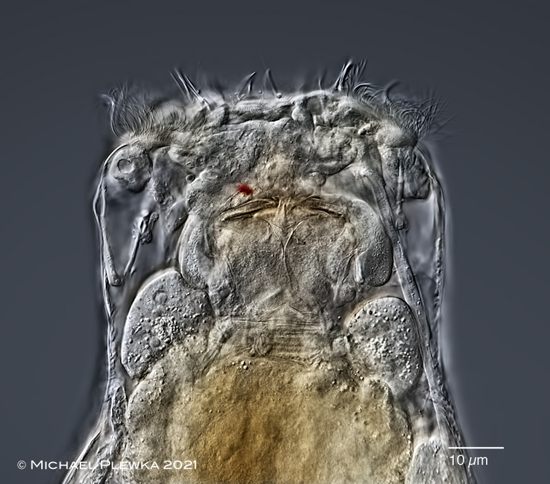 |
| Enteroplea lacustris, same specimen anterior part; focus plane on the mastax and gastric glands. Also visible is the single cerebral eyespot. (2) |
| |
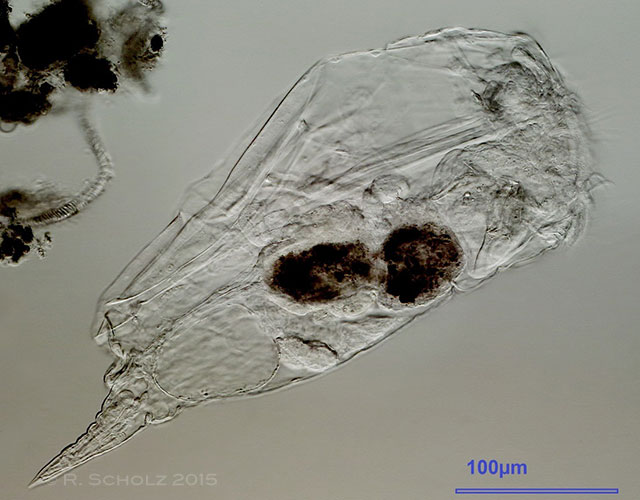 |
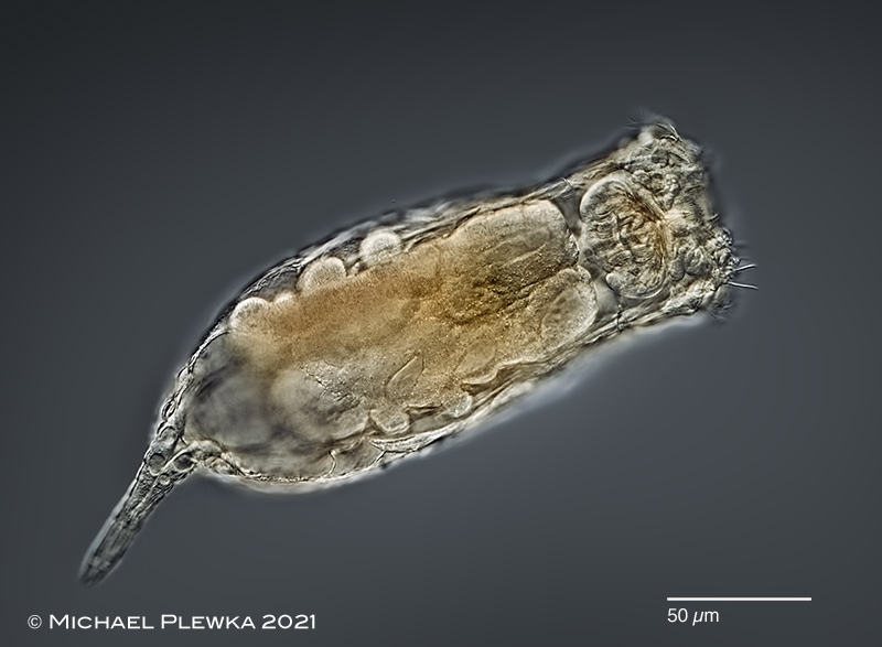
| Enteroplea lacustris, lateral view, slightly compressed specimen. (1) |
| |
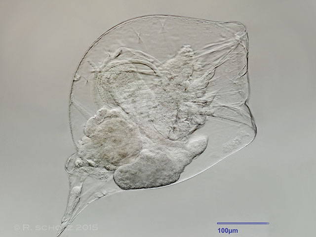 |
| Enteroplea lacustris; head completely retracted. The transparent integument reveals a perfect view of the pseudocoel. (1) |
| |
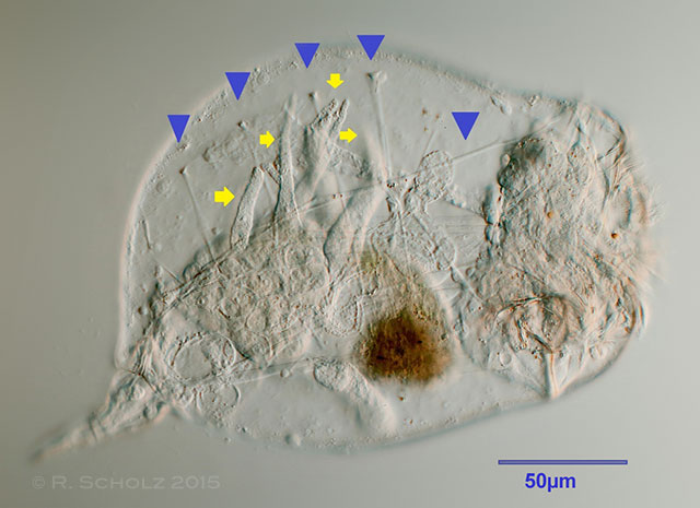 |
| Enteroplea lacustris, gastric glands with typical appendages (yellow arrows). The blue arrowheads point to the insertion points of some transversal muscle fibers. (1) |
| |
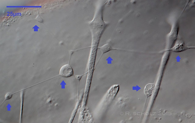 |
| Enteroplea lacustris, amoeboid tissue (blue arrows). (1) |
| |
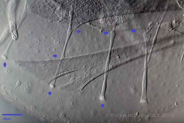 |
| Enteroplea lacustris; as some of the filaments seem to be connected to muscle fibers, they could also be part of the nervous system (?). (1) |
| |
|
|
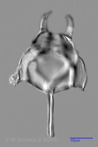
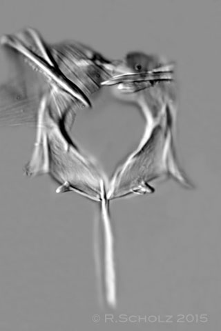
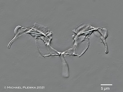
| Enteroplea lacustris; three aspects of the trophi (stacked images) (1) |
| |
  |
| |
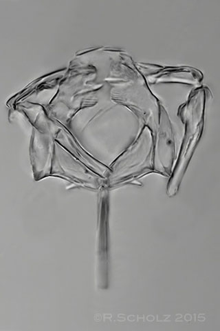
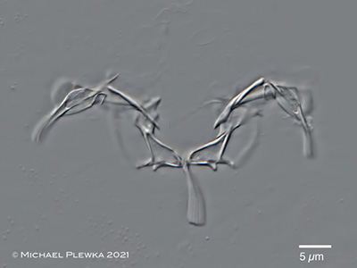
| Enteroplea lacustris; 2 aspects of trophi of specimen from (2) |
| |
| |
| |
| |
| |
| Location (1): “Rotes Moor” nearby the city of Wesenberg/Mecklenburg-Vorpommern/Germany |
Habitat (1): “Bruch” with waterplants, together with Cephalodella gigantea. About one specimen
per ml. Conductance: 85µS, pH: 6.5. |
| Date (1):collected 14.7.2015, images: 22.7.2015 |
| Images of sample (1) courtesy of Richard Scholz. |
| |
| |
| |
|
Location (2): Fondotoce, Verbania, Piemont, Italy; pond |
 |
| |
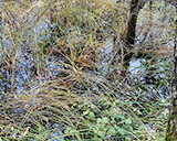
| Habitat (2): periphyton (click to enlarge >>>>) |
| |
| Date (2): 08.10.2021 |
| |
|
|
|
|
|
| |
| |
|
|